Hair Density Analysis
Measure scalp coverage with calibrated hairs-per-area density mapping.
Try It Free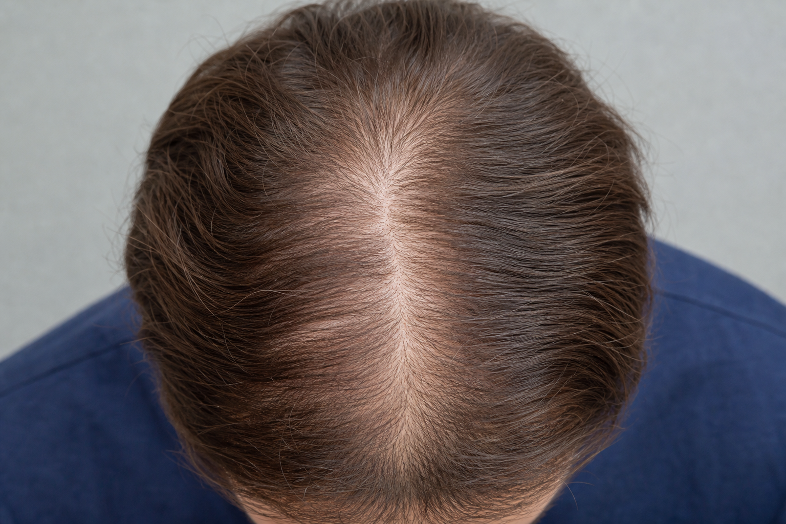
Overview
Density analysis goes beyond raw counts by calculating hairs per square centimeter (or mm²) across the scanned region. Enter your image scale or use our calibration tools to get clinical-grade density metrics used in transplant planning and hair loss staging.
What You Get
Key Features
- Automatic hairs-per-cm² and hairs-per-mm² calculation
- Compare density across different scalp regions
- Assess donor area suitability for transplant procedures
- Track density improvements during treatment programs
- Export density metrics for patient records and reports
- Combine with visit tracking for longitudinal charts
Who Uses It
Transplant Surgeons
Evaluate donor density before harvesting and plan graft counts.
Dermatologists
Objectively measure whether topical or oral treatments increase density.
Hair Restoration Centers
Show patients measurable before/after density improvements.
Research Trials
Standardize density endpoints across multi-site clinical studies.
Image Guidelines
- Provide accurate image scale (cm or mm per pixel) for calibration
- Capture a consistent area size across follow-up visits
- Use the same scalp location each time for valid comparisons
- Avoid overlapping hair that obscures the scalp surface
Related Analyses
 CORE
CORE
AI strand detection
Hair Counting
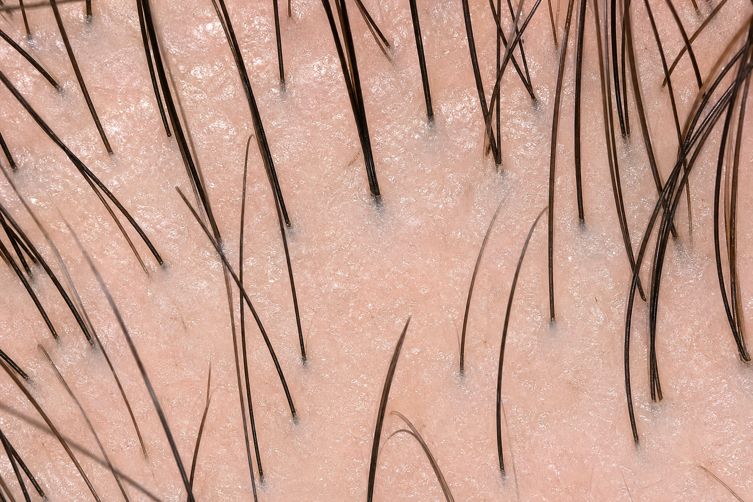
Our core hair counting model detects every visible strand in micro-camera or dermatoscope images. It groups hairs into follicular units (1–7 hairs) and returns an annotated image with confidence scores — the foundation for density tracking, transplant planning, and treatment monitoring.
Learn more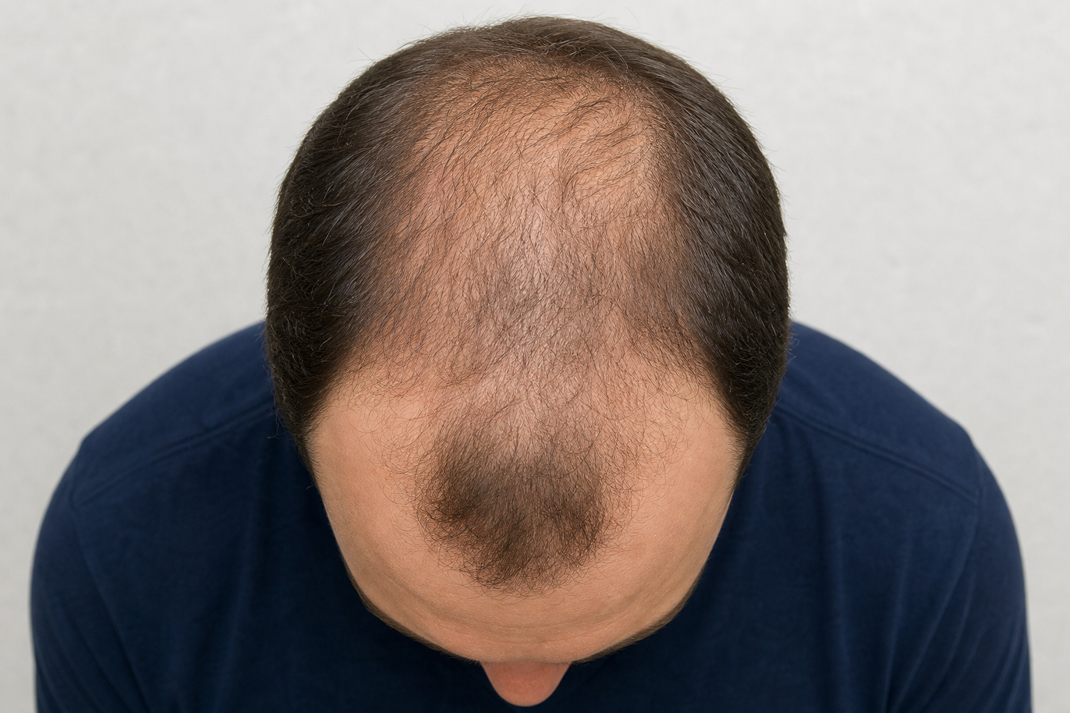 DIAGNOSTIC
Norwood staging
Baldness Analysis
Upload a top-down scalp photo and our AI maps hair loss zones, estimates the Norwood–Hamilton classification stage, and highlights affected areas. Ideal for initial consultations, progress tracking, and patient education about pattern baldness severity.
Learn more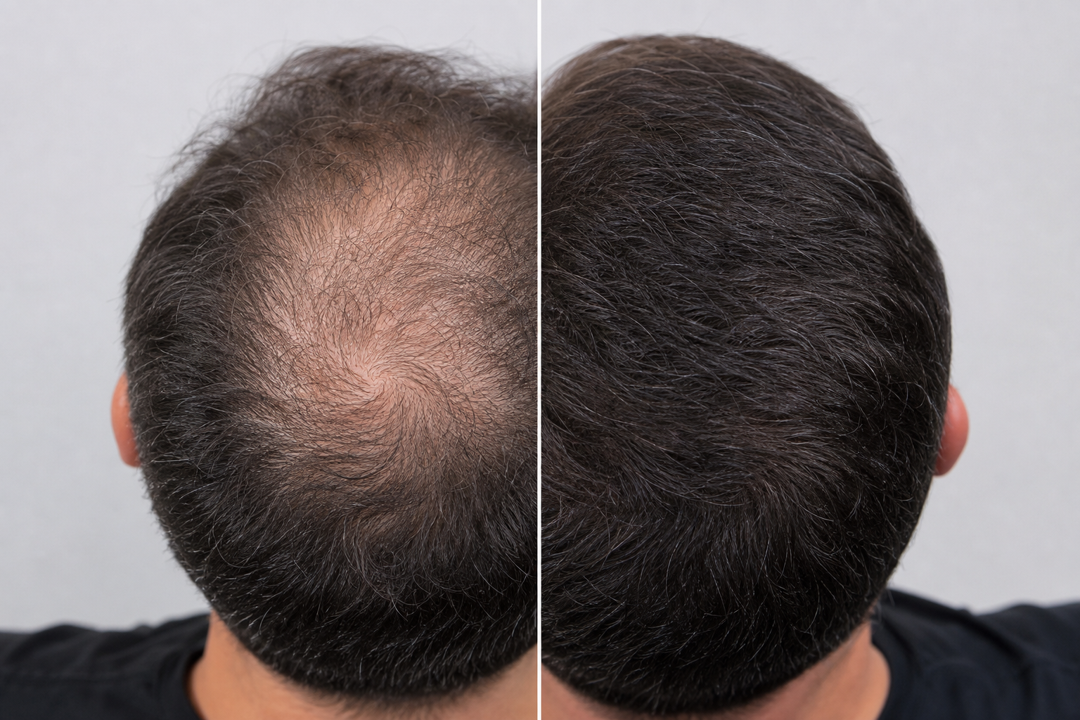 PROGRESS
Treatment tracking
Before & After
Upload two scalp images taken at different times and get an AI-powered side-by-side comparison. See density changes, Norwood stage shifts, and improvement percentages — perfect for demonstrating treatment efficacy to patients or documenting transplant outcomes.
Learn moreReady to get started?
Try Density Analysis free — no credit card required.